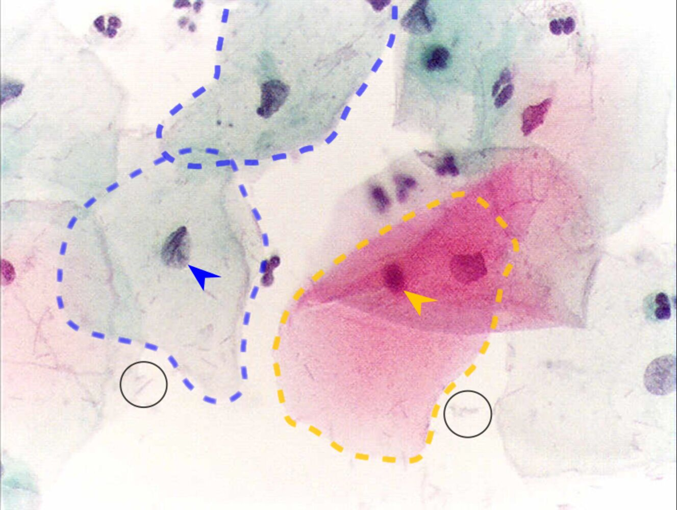
 Normal squamous epithelial cells are present.
- The cells with blue (cyanophilic) cytoplasm on the left of the image are intermediate cells (marked by blue dotted line).
- The cell with the red (eosinophilic) cytoplasm on the right of the image is a superficial cell (marked by yellow dotted line).
- Neutrophils can also be seen (small cells with a segmented nucleus).
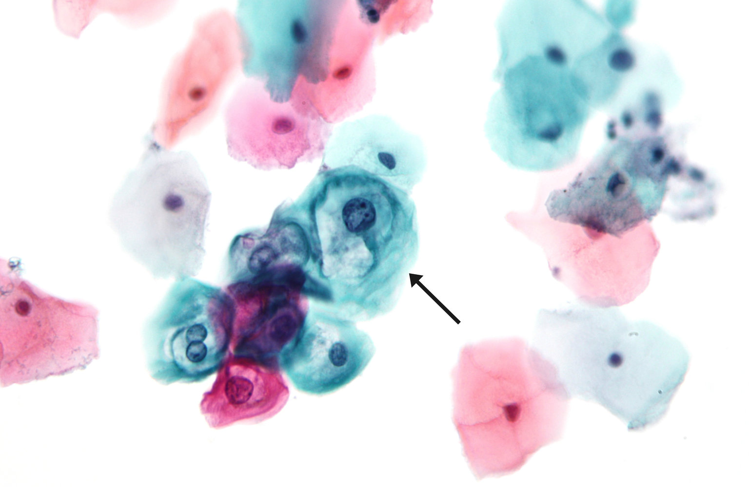 Pap smear showing low-grade dysplasia
In the center of the image is an irregular clump of koilocytes (marked by an arrow). The koilocytes are characterized by perinuclear halos and show slight nuclear enlargement with irregularly distributed chromatin; they may also exhibit binucleation.
Pap smear showing low-grade dysplasia
In the center of the image is an irregular clump of koilocytes (marked by an arrow). The koilocytes are characterized by perinuclear halos and show slight nuclear enlargement with irregularly distributed chromatin; they may also exhibit binucleation.
Tip
Koilocytes are epithelial cells with perinuclear halos that are pathognomonic of HPV infection and may be present from early HPV infection.
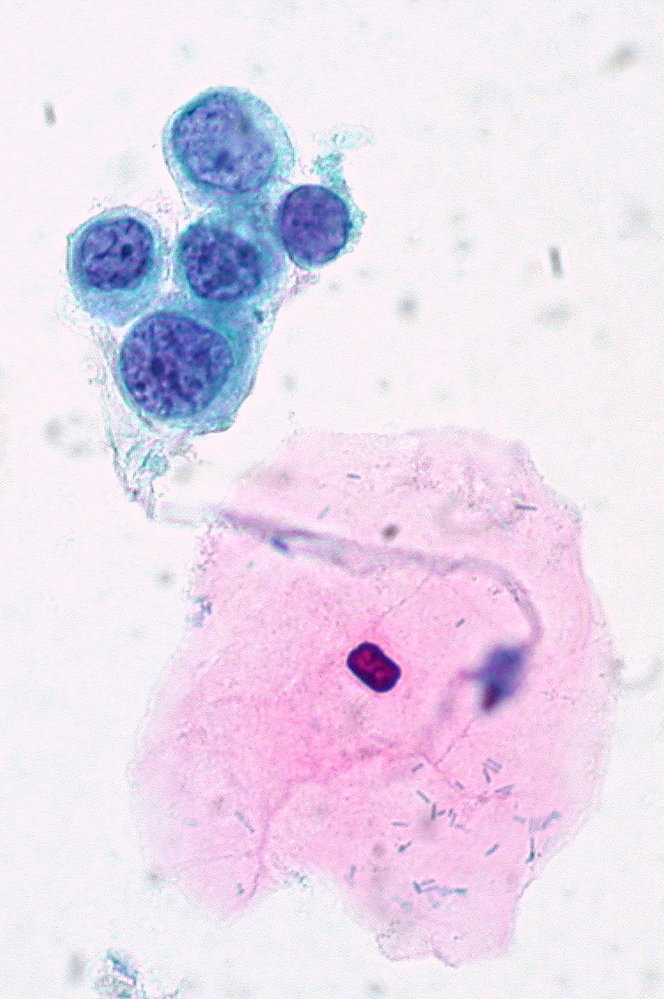 Pap smear showing severe dysplasia
There is a loosely connected group of abnormal squamous epithelial cells (blue-green cytoplasm). The nuclei are variable in size with a high nuclear-cytoplasmic ratio and there are course clumps of chromatin. The large cell with pink cytoplasm is a normal superficial squamous cell.
Pap smear showing severe dysplasia
There is a loosely connected group of abnormal squamous epithelial cells (blue-green cytoplasm). The nuclei are variable in size with a high nuclear-cytoplasmic ratio and there are course clumps of chromatin. The large cell with pink cytoplasm is a normal superficial squamous cell.