Epidemiology
Etiology
CJD is caused by misfolded proteins (prions, PrPSc) that are either produced by affected individual themselves, or taken up from an exogenous source.
- Sporadic (∼ 85%): no identifiable source
- Familial (∼ 10–15%)
- Acquired (< 1%)
- Iatrogenic CJD: transmission during medical procedures, such as:
- Brain surgery (surgical equipment)
- Organ transplantation (e.g., corneal transplant)
- Blood transfusion
- Variant CJD (vCJD)
- Occurs due to ingestion of beef infected with bovine spongiform encephalopathy (BSE)
- BSE is a transmissible prion disease occurring in cattle also known as “mad cow disease”
- Iatrogenic CJD: transmission during medical procedures, such as:
Pathophysiology
- Conversion of normal cellular prion proteins with alpha-helical structure (PrPc) to prions that demonstrate an increase in beta-pleated sheet structure (PrPSc) → conformational change of physiological PrPc → PrPSc accumulation and plaque formation → neuronal cell death → progression to spongiform encephalopathy
- Conformational change is triggered via misfolded PrPSc (from scrapie, a transmissible spongiform encephalopathy of sheep)
- Since misfolded prions are insoluble, they deposit as plaques resistant to proteases and standard autoclaving, thus contributing to the formation of more PrPSc.
Clinical features
- Neurological symptoms
- Cerebellar disturbances (e.g., gait instability) and extrapyramidal deficits
- Myoclonus
- A sudden involuntary twitch of a muscle or groups of muscles caused by muscular contraction or inhibition
- Often triggered by startling (e.g., loud noises)
- Also associated with metabolic abnormalities found in liver and renal failure
- Ataxia
- Neuropsychiatric symptoms
- Rapidly progressing dementia (weeks to months)
- Personality changes
- Akinetic mutism
Tip
Rapidly progressive dementia and myoclonic jerks are the hallmarks of Creutzfeldt-Jakob disease.
Diagnostics
- CSF analysis
- ↑ 14-3-3 protein
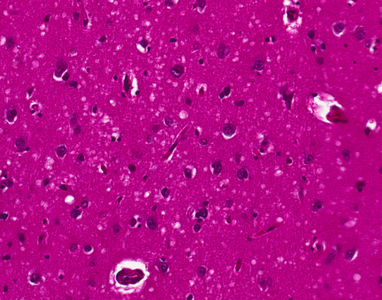
- Brain biopsy
- Diagnosis can only be confirmed by biopsy/autopsy and subsequent neuropathological examination.
- Microscopic findings include spongiform degeneration (e.g., intracytoplasmic vacuoles within the neurons of cerebral and cerebellar cortex that can be seen on H&E)