| Receptor | Change in Second Messenger | Primary Effects |
|---|---|---|
| Alpha-1 | ↑ IP3 | Peripheral vasoconstriction Urethral constriction Pupillary dilation |
| Alpha-2 | ↓ cAMP | CNS sympatholytic ↓ Insulin release & intestinal motility |
| Beta-1 | ↑ cAMP | ↑ Cardiac contractility & heart rate ↑ Renin release by JG cells of kidney |
| Beta-2 | ↑ cAMP | Peripheral vasodilation Bronchodilation |
| Muscarinic-2 | ↓ cAMP | ↓ Cardiac contractility & heart rate |
| Muscarinic-3 | ↑ IP3 | Bronchoconstriction ↑ Insulin release & intestinal motility Bladder contraction Pupillary constriction Peripheral vasodilation* |
Receptors
- Receptor Types
- Intracellular receptors
- Examples of ligands: Glucocorticoids
- Cell surface receptors
- G protein-coupled receptors
- Examples of ligands: Catecholamines
- Receptor tyrosine kinases
- Examples of ligands: Insulin
- Receptors with associated kinases
- Examples of ligands: Growth hormones
- Receptor protein serine/threonine kinases
- Examples of ligands: TGF-β (cytokine)
- Ligand-gated ion channels
- Examples of ligands: Acetylcholine
- G protein-coupled receptors
- Intracellular receptors
Receptor tyrosine kinases (RTKs)
- Examples of ligands: insulin, growth factors (e.g., EGF, IGF, FGF, TGF)
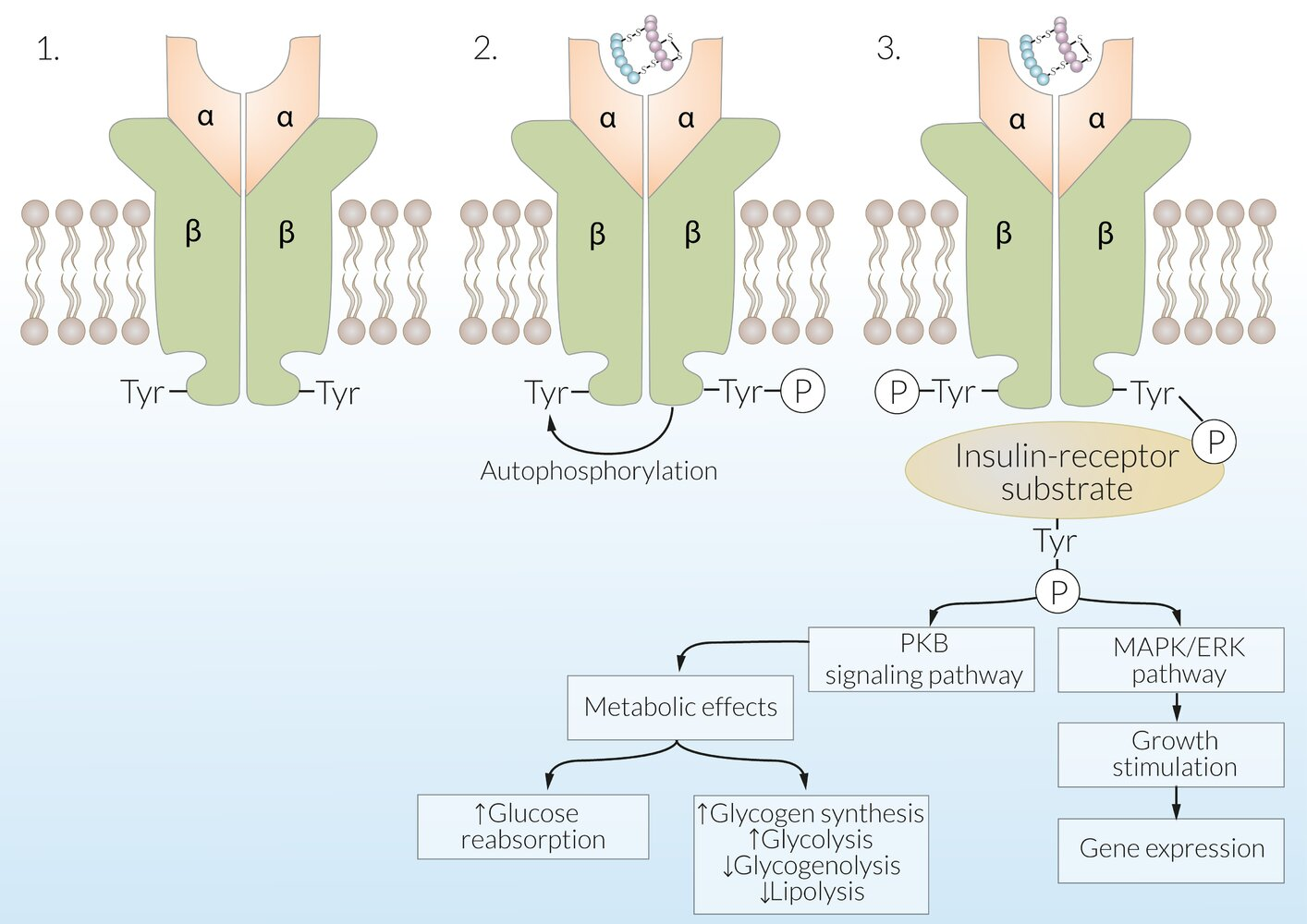
- Activation of tyrosine kinase → ↓ cAMP → activation of protein phosphatase 1 (PP 1) → PP 1-mediated dephosphorylation deactivates glycogen phosphorylase and activates glycogen synthase → ↓ glycogenolysis and ↑ glycogen synthesis
Non-receptor tyrosine kinases
- Examples of ligands: growth hormone, prolactin, erythropoietin, thrombopoietin, cytokines (e.g., G-CSF, IFN, IL-2, IL-6)
- Activation principle (similar to receptor tyrosine kinases)
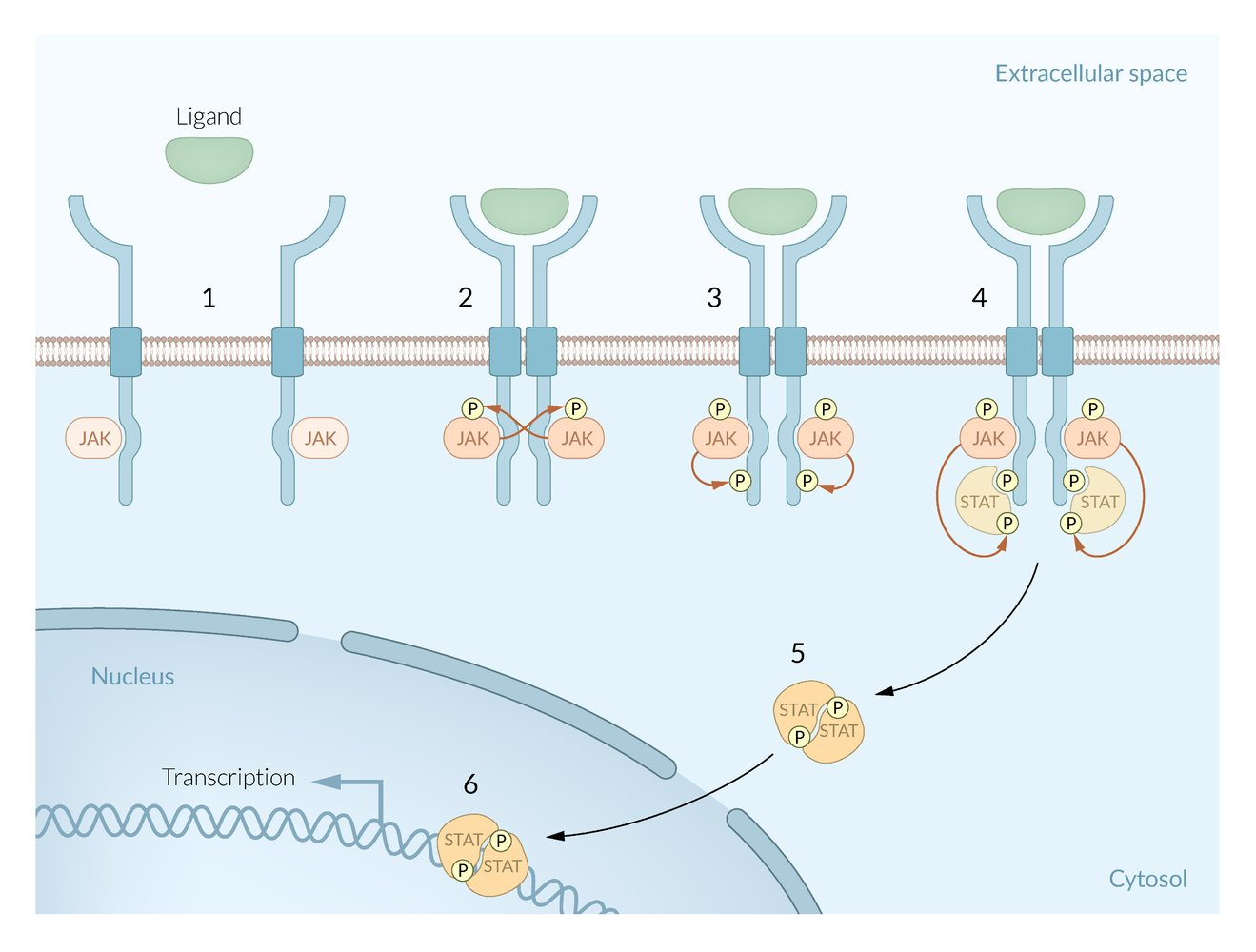
- Ligand binding leads to receptor dimerization.
- Two neighboring tyrosine kinase domains of JAK phosphorylate each other (autophosphorylation) → JAK activation
- Development (at the phosphorylated tyrosine residues) of binding sites for SH2 domains of signal proteins (STAT proteins).
- STAT proteins are phosphorylated and dimerized by JAK.
- STAT dimers exert their effect directly in the nucleus as a transcription factor for JAK-STAT regulated genes.
Tip
- JAK-STAT activation is related with 3 types of Myeloproliferative neoplasms
- JAK-STAT deficiency is related with
Ligand-gated ion channels
- Since most ligands of ligand-gated ion channels are neurotransmitters, only a short overview is provided here.
- Examples of ligands: acetylcholine, GABAA, glutamate, IP3
- Receptors: Nicotinic Acetylcholine Receptors (nAChRs), GABA/Glycine Receptors, Glutamate Receptors
- Receptor structure: Cell surface receptors that act as ion channels
- Activation principle (example)
- Ligand binding → conformational change
- Pore opening enables ion movement (Na+, K+ Ca2+, Cl-) across the cell membrane.
- Altered membrane potential → rapid signal response
Second messengers
cAMP (cyclic adenosine monophosphate) and protein kinase A
- Regulation of adenylyl cyclase: depends on the type of G protein of the G protein-coupled receptor
- Gs proteins: activate adenylyl cyclase → ↑ cAMP
- Gi proteins: inhibit adenylyl cyclase → ↓ cAMP
- Effects
- cAMP activates protein kinase A (PKA).
- Mechanism: PKA controls the activity (activation or inactivation) of numerous enzymes via phosphorylation of serine and threonine residues.
- Example: glycogen metabolism to ↑ blood glucose concentration
- Hypoglycemia → ↑ glucagon or adrenaline → GPCR activation → ↑ cAMP → activation of PKA, which causes:
- Phosphorylation of glycogen phosphorylase → activation → increased mobilization of glucose from glycogen
- Phosphorylation of glycogen synthase → inhibition→ decreased glycogen formation
- Hypoglycemia → ↑ glucagon or adrenaline → GPCR activation → ↑ cAMP → activation of PKA, which causes:
- Degradation
- cAMP is degraded by phosphodiesterase to adenosine monophosphate (AMP).
- Examples of ligand hormones
- Hypothalamic hormones: GHRH, CRH, vasopressin (V2 receptor)
- Pituitary hormones: TSH, ACTH, FSH, LH, MSH
- Other hormones: PTH, calcitonin, glucagon, histamine (H2 receptor), hCG
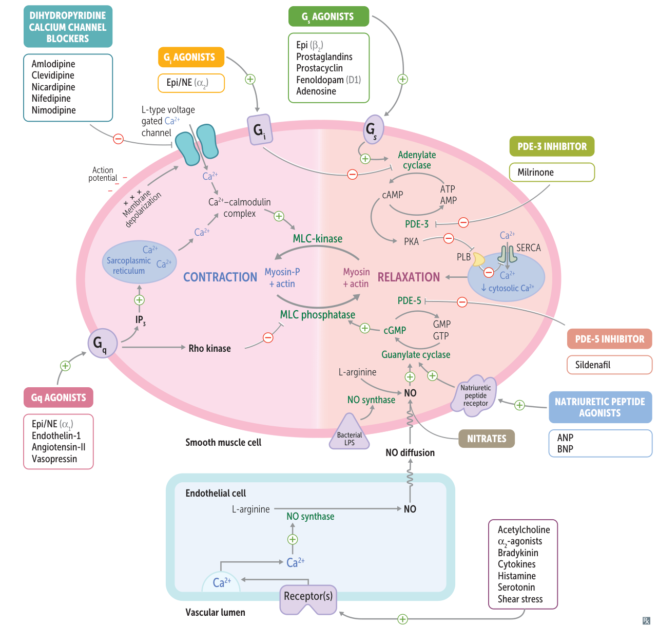
cGMP (cyclic guanosine monophosphate)
- Synthesis: cGMP is synthesized from GTP by the guanylate cyclase. There are two subforms of guanylate cyclase:
- Soluble guanylate cyclase: activated by nitric oxide
- Membrane-bound guanylate cyclase: activated through extracellular ligand binding (e.g., EDRF, ANP, BNP)
Tip
Both lead to vasodilation.
- Effects
- Activates cGMP-dependent protein kinase G in smooth muscle cells → inhibits Ca2+ outflow from the sarcoplasmic reticulum → ↓ intracellular Ca2+ → relaxed smooth vascular muscles → vasodilation
- cGMP-dependent ion channels in the photoreceptor cells of the retina → preserve the unstimulated state → dark signal
- Degradation: by phosphodiesterase to GMP
- So PDE inhibitors work as vasodilators, used in the treatment of pulmonary hypertension (PDE-4 inhibitor roflumilast) and erectile dysfunction (PDE-5 inhibitor sildenafil).
IP3 and DAG
- Synthesis
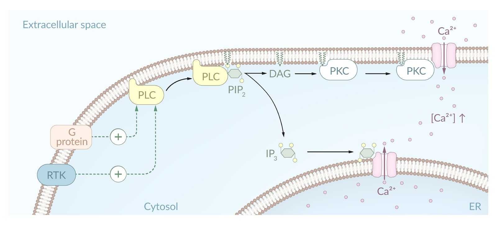
- Activation of a Gq protein-coupled receptor
- Activation of phospholipase C (an enzyme that cleaves phospholipids)
- Cleaving of the membrane-bound phospholipids PIP2 (phosphatidylinositol 4,5-bisphosphate) into the second messenger IP3 (inositol triphosphate) and DAG (diacylglycerol)
- Function
- IP3 (hydrophilic) diffuses into the cytoplasm → activation of IP3 receptors at the membrane of the endoplasmic reticulum (ER) → Ca2+ release from the ER via the IP3 receptor-coupled calcium channel → ↑ intracellular Ca2+ concentration → smooth muscle contraction
- DAG (lipophilic) remains in the membrane → activates protein kinase C (PKC)
- PKC is Ca2+-dependent (depends on IP3-mediated Ca2+ release)
- Mechanism of action: regulates the activity of various enzymes via phosphorylation of serine and threonine residues, e.g., regulatory proteins that influence cell growth (actin cytoskeleton) or differentiation (EGFR)
- Examples of the effect of PKC: cell growth and proliferation
- Examples of ligands: vasopressin (V1 receptor), oxytocin, GnRH, TRH, angiotensin, gastrin, histamine (H1 receptor)
Ca2+ as a second messengers
- Effect of Ca2+, especially as a complex with calmodulin: One molecule of calmodulin binds up to four Ca2+ ions → conformational change → regulation of calmodulin-dependent kinases/phosphatases, e.g., CaM kinase III (protein synthesis)