Epidemiology
Etiology
- Pathogen: Mycobacterium leprae is an obligate, intracellular, acid-fast bacillus that cannot be cultured and thrives in cold temperatures.
- Route of transmission
- Close contact with fomites, contaminated soil, infected individuals, and nine-banded armadillos (in rare cases)
- Respiratory droplet transmission
- Risk factors are close contact with infected individuals or contaminated soil.
Pathophysiology
- Disease spectrum depends on the host’s cell-mediated immune response (Th1 vs. Th2).
- Tuberculoid Leprosy (Paucibacillary)
- Strong Th1 Response (High IL-2, IFN-γ).
- Macrophage activation limits bacterial growth.
- Low bacterial load.
- (+) Lepromin skin test (indicates strong immune response).
- Histology: Non-caseating granulomas.
- Lepromatous Leprosy (Multibacillary)
- Weak Th1 / Strong Th2 Response (High IL-4, IL-5, IL-10).
- Depressed cell-mediated immunity; humoral response is ineffective.
- High bacterial load.
- (-) Lepromin skin test (anergy).
- Histology: Foamy histiocytes (macrophages) packed with AFB (Virchow cells); no granulomas.
Clinical features
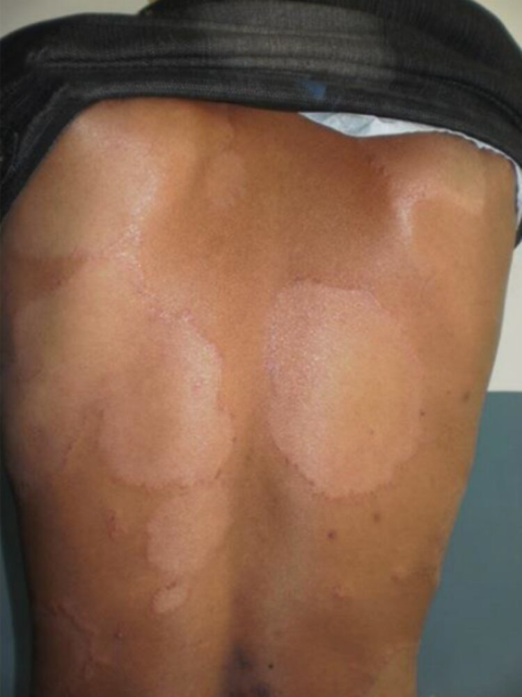
- Tuberculoid Form:
- Hypopigmented, hairless skin plaques.
- Focal loss of sensation (anesthesia) at the lesion site.
- Enlarged peripheral nerves (palpable).
- Lepromatous Form:
- Diffuse nodular lesions on extensor surfaces and face.
- Leonine facies (thickening of facial skin, loss of eyebrows/eyelashes).
- Saddle nose deformity (septal perforation).
- Glove and stocking peripheral neuropathy (sensory loss > motor).
- Testicular destruction (infertility/gynecomastia).
Diagnostics
The lepromin skin test (in which M leprae antigens are injected intradermally) can be used to distinguish between tuberculoid and lepromatous leprosy. Patients with tuberculoid leprosy will develop an indurated nodule at the site of the injection (much like a positive PPD test for M tuberculosis). In contrast, the test is usually nonreactive in patients with lepromatous leprosy due to their weak TH1 cell-mediated immune response.
Treatment
Dapsone
- Mechanism of action
- Competitive antagonist of para-aminobenzoic acid (PABA) for dihydropteroate synthetase → inhibition of dihydrofolic acid synthesis
- Structurally different from sulfonamides but a similar mechanism of action
- Clinical use
- M. leprae: lepromatous and tuberculoid leprosy
- P. jiroveci pneumonia
- Prophylaxis
- Treatment: used in combination with TMP as an alternative to TMP/SMX
- Adverse effects
- Methemoglobinemia
- Triggers hemolytic anemia in patients with G6PD deficiency
- Agranulocytosis