Epidemiology
Etiology
- Nontraumatic (spontaneous)
- Hypertension: most common cause of spontaneous ICH
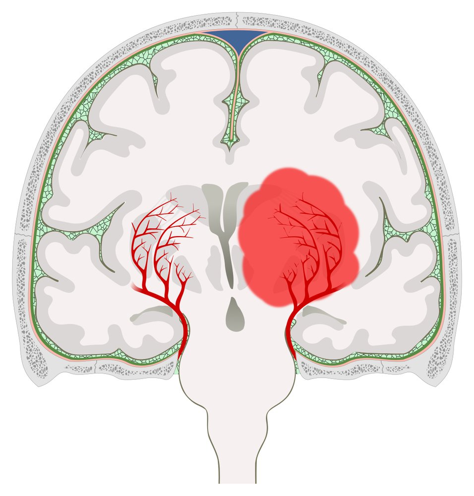
- Hypertension-induced hemorrhages typically occur in the subcortical regions. Small penetrating arteries supply this part of the brain and emerge at sharp angles from larger vessels, making them particularly sensitive to changes in blood pressure.
- Cerebral amyloid angiopathy: most common cause of spontaneous ICH in individuals > 60 years of age
- Typically causes lobar hemorrhage
- Arteriovenous malformations: most common cause of spontaneous intracerebral hemorrhage in children
- Vasculitis (e.g., giant cell arteritis)
- Neoplasms (e.g., meningioma)
- Ischemic stroke (due to reperfusion injury)
- Hypertension: most common cause of spontaneous ICH
Pathophysiology
- Nontraumatic mechanisms of hemorrhage
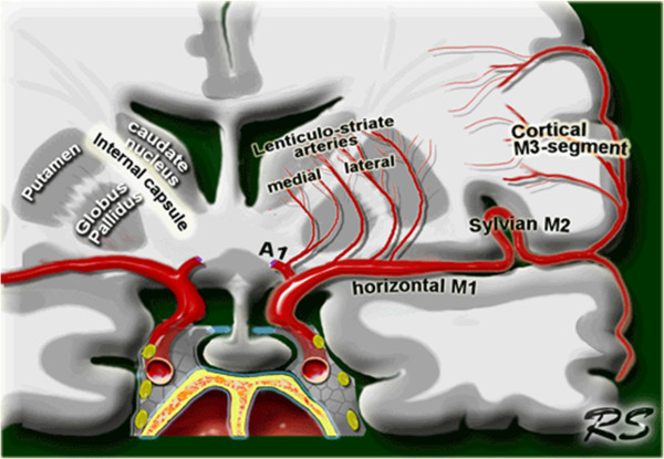
- Chronic arterial hypertension → lipohyalinosis of lenticulostriate vessels (which supply the basal ganglia) and/or formation and rupture of Charcot-Bouchard microaneurysms → lacunar strokes (ischemia) of the basal ganglia. See also Ischemic stroke > Lacunar infarction
- Putamen most commonly affected, which is supplied by lenticulostriate arteries
Charcot-Bouchard aneurysms vs Saccular (berry) aneurysms
Feature Charcot-Bouchard Microaneurysms Saccular (Berry) Aneurysms Associated Conditions Hypertension ADPKD, Ehlers-Danlos syndrome, hypertension Location Basal ganglia
Cerebellum
Thalamus
PonsCircle of Willis Size <1 mm Variable, 2-25 mm Result of Rupture Intracerebral hemorrhage Subarachnoid hemorrhage Symptoms of Rupture Progressive neurologic deficits
Headache may followSudden severe headache
Focal neurologic deficits uncommon-aneurysms/../../../appendix/Pasted-image-20250304102040.png)
-aneurysms/../../../appendix/Pasted-image-20240526113641.png)
- Saccular: This term means “resembling a sac.” Saccular aneurysms are outpouchings or bulges on one side of a blood vessel wall.
- Berry: The “berry” description refers to the characteristic round shape of these aneurysms. They look like a berry connected to the main artery.
- Other locations: thalamus (second most common) and infratentorial parts of the brain (e.g., pons, cerebellum)
- Putamen most commonly affected, which is supplied by lenticulostriate arteries
- Cerebral amyloid angiopathy: deposition of β-amyloid peptides in vessel walls → focal damage with formation of microaneurysms → rupture → recurrent lobar intracerebral hemorrhage
- Chronic arterial hypertension → lipohyalinosis of lenticulostriate vessels (which supply the basal ganglia) and/or formation and rupture of Charcot-Bouchard microaneurysms → lacunar strokes (ischemia) of the basal ganglia. See also Ischemic stroke > Lacunar infarction
- Traumatic: blunt or penetrating injury → damage to vessels
Clinical features
- Headache
- Focal neurologic signs and symptoms
- Putaminal hemorrhage: contralateral hemiparesis or hemiplegia with less severe contralateral hemisensory loss; eyes deviate toward the side of the hematoma
- Thalamic hemorrhage: contralateral hemiparesis, contralateral hemisensory loss, decreased consciousness, wrong way eyes
- Putaminal hemorrhage: contralateral hemiparesis or hemiplegia with less severe contralateral hemisensory loss; eyes deviate toward the side of the hematoma
Mnemonic
- The dorsal striatum is composed of both the caudate and the putamen.
- The caudate is involved in both motor activity as well as more cognitive functions, while the putamen is primarily involved in motor activity.
- The caudate nucleus is the most caugnitive part of the basal ganglia.
- When you see put-am-en on an exam, “put-an-em” for Motor.
- Course
- Symptoms typically progress gradually over minutes to a few hours
- Focal deficits worsen with expansion of the hematoma
- Late: symptoms of increased ICP
- Nausea and vomiting
- Confusion and loss of consciousness
- Bradycardia
- Fixed pupils