Meningiomas are a diverse group of brain tumors that arise from the arachnoid layer (specifically the arachnoid cap cells) and can therefore occur in any part of the CNS with a meningeal covering.
Epidemiology
- Most common benign primary brain tumor in adults
Etiology
- Multiple meningiomas may develop in patients with neurofibromatosis type II.
Pathophysiology
Clinical features
- Mostly asymptomatic
- Meningiomas are slow-growing tumors and, as such, they are less invasive and the body has more time to adapt to the tumor.
- General symptoms of CNS tumors (e.g., seizures and focal neurologic signs)
Diagnostics
MRI (imaging modality of choice)
- Plain MRI findings
- Round, sharply demarcated space-occupying lesion with radiological features of an extra-axial tumor
- Dural tail sign
- T1: isointense or hypointense
- T2: isointense or hyperintense
- Contrast MRI findings
- Significant homogenous enhancement of the meningioma
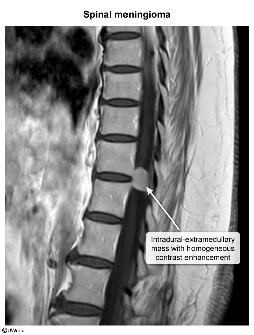
- As meningiomas grow, they often stimulate angiogenesis
- Significant homogenous enhancement of the meningioma
Pathology
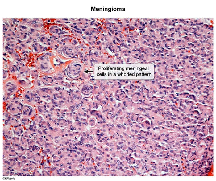
Microscopic findings
- Mesenchymal origin (arachnoid cap cells)
- Whorls of meningothelial cells (onion peel arrangement)
- Psammoma bodies
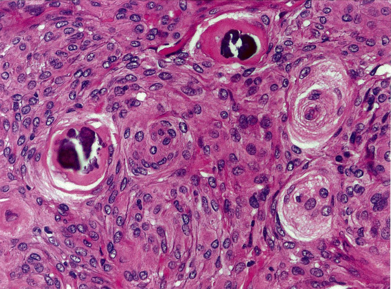
 Two of these whorls have a deeply basophilic acellular core, known as a psammoma body, which indicates dystrophic calcification.