Etiology
Pathogen
- Gram-positive, filamentous, branching rods.
- Anaerobe (contrast with Nocardia, which is aerobic).
- Not acid-fast (contrast with Nocardia, which is weakly acid-fast).
- Normal flora of the oral cavity, reproductive tract, and GI tract.
Pathophysiology
- Infection usually follows mucosal trauma (e.g., dental procedures, surgery) allowing invasion of deep tissue.
- Spreads contiguously via slow-growing, indurated masses.
- Forms abscesses with draining sinus tracts through the skin.
Clinical features
- Cervicofacial Actinomycosis:
- Most common form.
- Associated with dental trauma/extraction or poor oral hygiene.
- Presents as a firm, nontender lump on the jaw (“lumpy jaw”) that progresses to abscesses with sinus tracts.
- Becomes indurated with purulent discharge that contains sulfur granules from fistulae and draining sinus tracts.
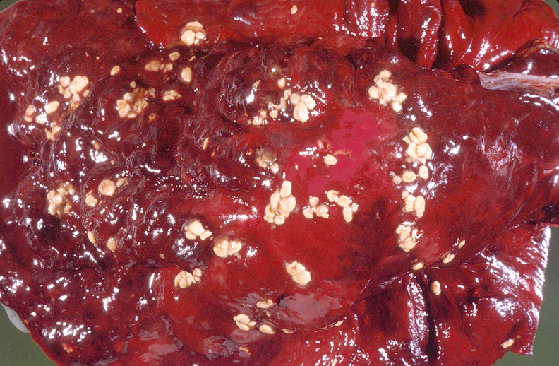
- Sulfur granules refer to macroscopic grains – approx. 1 mm in diameter – of hard clumps of bacterial filaments, pus, debris, and hyaline. The granules appear yellow within pus, although despite the name they do not contain sulfur.
- Pelvic Actinomycosis:
- Strongly associated with long-term use of Intrauterine Devices (IUDs).
- Thoracic/Abdominal:
- Thoracic: Aspiration (can be confused with lung malignancy).
- Abdominal: Post-surgery (e.g., appendicitis).
Tip
Definitive diagnosis is based on the identification of actinomycotic sulfur granules or bacteria.
Diagnostics
- Microscopy: direct visualization and staining of specimen → accumulations of radially protruding and branching Actinomyces (conglomerates with a “cauliflower-like” appearance) that are surrounded by numerous granulocytes
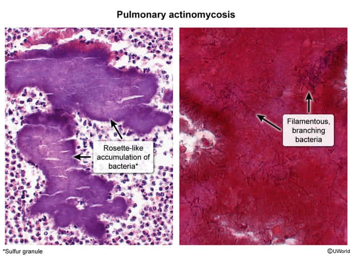

An aggregate of basophilic bacteria with radially branching filaments is visible at the center, which has a cauliflower-like appearance (yellow overlay).
The bacteria are surrounded by numerous granulocytes, resulting in a lesion with a rosette-like pattern (green overlay).
This is the typical histopathological appearance of a yellow “sulfur granule” caused by actinomycosis.
Treatment
Penicillin t