Peak incidence: 70–79 years (rarely seen in patients < 50 years)
Etiology
Association with polymyalgia rheumatica (PMR): up to 50% of patients with giant cell arteritis also have PMR.
Pathophysiology
Monocytes differentiate into macrophages and giant cells, which produce cytokines (e.g., IL-6, TNF-α) that augment the inflammatory response → focal granulomatous inflammation
Most commonly involves external carotid artery branches (especially temporal artery), as well as the aorta and vertebral arteries
Clinical features
Cranial giant cell arteritis: involves the extracranial branches of the common carotid, internal carotid, and external carotid arteries (the temporal artery is the most commonly affected vessel)
New-onset unilateral (or bilateral) headache
Can be pulse-synchronous, throbbing, dull
Typically located over the temples
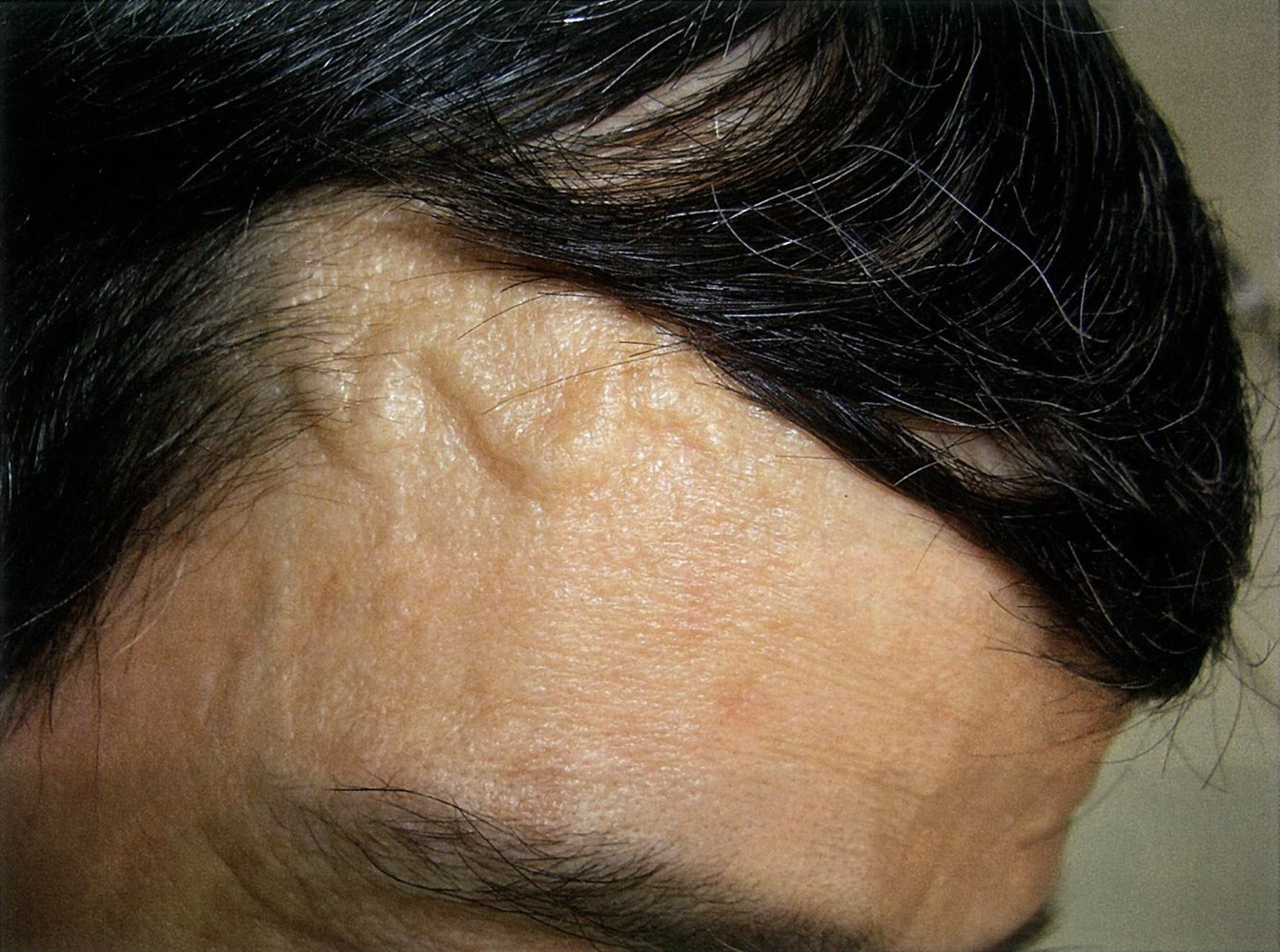
Hardened and tender temporal artery
Craniofacial pain syndromes: Jaw claudication, tongue claudication, and facial pain may occur. This tends to appear during mastication (chewing) when the blood supply to the corresponding areas does not increase normally due to the narrowing of the arterial lumens.
Sudden vision loss: due to inflammation and occlusion of the ophthalmic artery and its branches