A gram-positive, nonsporulating, club-shaped bacillus
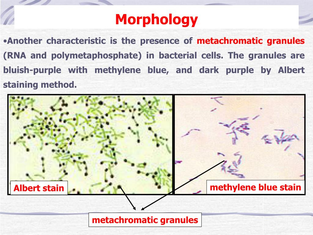
Contains metachromatic granules (volutin granules; stain red with a blue dye)
Route of infection
Droplet transmission
Pathophysiology
C. diphtheriae has both toxigenic and nontoxigenic strains; toxigenic strains contain a beta-prophage gene (tox), which encodes for the exotoxin diphtheria toxin
Conversion from nontoxigenic to toxigenic C diphtheriae occurs due to infection with a lysogenic bacteriophage called Corynephage beta.
This phage inserts the tox gene into the C diphtheriae genome, which results in the bacterial expression of the diphtheria AB toxin.
Diphtheria toxin irreversibly halts protein synthesis due to ADP-ribosylation of elongation factor-2 and causes severe local (eg, pseudomembranous pharyngitis) and systemic (eg, myocarditis, neuritis) effects.
Clinical features
Local features
Tonsillar and pharyngeal diphtheria
Grayish-white pseudomembrane over the posterior pharyngeal wall, and/or tonsils. Also see Gray-white exudates of throat.
Any attempt to scrape off the pseudomembrane exposes the underlying capillaries and results in heavy bleeding.
Bull neck due to cervical lymphadenopathy and swelling of the soft tissue of the neck → airway obstruction