Epidemiology
Etiology
Pathophysiology
Progression
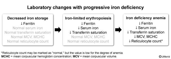
- Decreased iron storage: When there is a net negative iron balance, iron is first removed from the storage pool (eg, hepatocytes, reticuloendothelial system); iron is stored intracellularly within ferritin complexes. Small amount of ferritin is also found in the blood, and serum levels generally fluctuate in proportion to iron stores. Therefore, the body’s depletion of storage iron can be detected through a low serum ferritin level.
- Iron-limited erythropoiesis: As iron stores become depleted, serum iron levels decrease, followed by a compensatory rise in transferrin and a resultant decrease in transferrin saturation. During this phase, there is iron-limited erythropoiesis, in which newly made erythrocytes (ie, reticulocytes) are iron deficient and reticulocyte production may begin to be limited. Because mature erythrocytes have a slow turnover (ie, 120 days), most cells sampled during this stage still have normal morphology, and the total hemoglobin value may still be normal.
- Iron deficiency anemia: If iron deficiency continues, the last phase is frank iron deficiency anemia, which is reflected by a low hemoglobin level with microcytosis (low mean corpuscular volume MCV) and hypochromia (low mean corpuscular hemoglobin concentration MCHC) in most erythrocytes. Reticulocyte production decreases, resulting in an inappropriately low number of reticulocytes.
Clinical features
Diagnostics
- ↑ Platelet count (reactive thrombocytosis)
- Mechanism unknown
Differential diagnostics
| Iron deficiency anemia | Anemia of chronic disease | |
|---|---|---|
| Serum ferritin | ↓ | Normal or ↑ |
| Serum iron | ↓ | ↓ |
| Mean corpuscular volume | ↓ | Normal or ↓ |
| Transferrin (total iron-binding capacity) | ↑ | Normal or ↓ |
| Transferrin saturation | ↓ | ↓ |
| Erythropoietin | ↑ | Variable* |
| Hepcidin | ↓ | ↑ |
| C-reactive protein | Normal | ↑ |
| Serum cytokines | Normal | ↑ |