LH and FSH
- LH
- Female individuals: triggers ovulation
- Male individuals: stimulates testosterone synthesis in Leydig cells
- FSH: stimulates the maturation of germ cells in both male and female individuals
Androgen
- Four types of androgen: testosterone, dihydrotestosterone (DHT), androstenedione, dehydroepiandrosterone (DHEA)
- Androstenedione and dehydroepiandrosterone (DHEA) are substrate in testosterone and estrogen synthesis
- Testosterone stimulates increased protein synthesis by skeletal myocytes. It has been misused and abused by athletes, bodybuilders, weight lifters, and others to enhance athletic performance or physique
Testosterone synthesis
Testosterone synthesis takes place in the testicles.
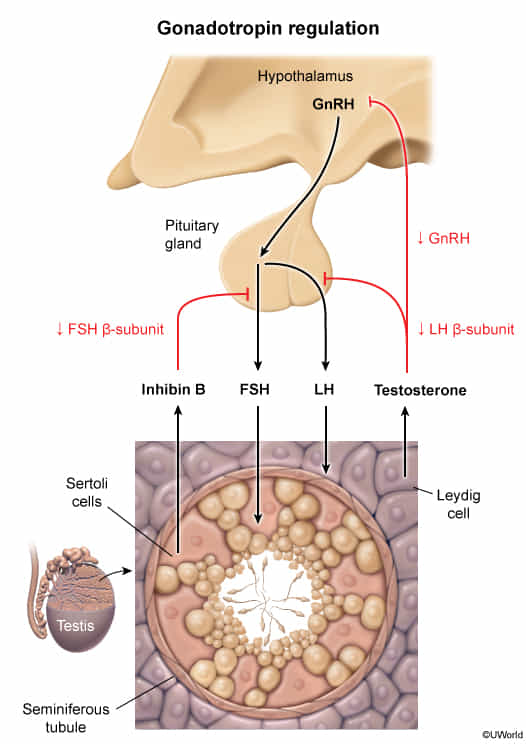
- The hypothalamus secretes gonadotropin-releasing hormone (GnRH) in pulses.
- GnRH stimulates the anterior pituitary cells to secrete luteinizing hormone (LH) and follicle-stimulating hormone (FSH).
- LH stimulates Leydig cells to produce testosterone.
- FSH stimulates Sertoli cells to release:
- Androgen-binding protein, stimulating spermatogenesis
- Inhibin B: Serves as negative feedback control for FSH secretion, a marker of Sertoli cell function and of spermatogenesis.

Mnemonic
- Sertoli cells are temperature sensitive, line seminiferous tubules, support sperm synthesis, and inhibit FSH
- Leydig cells secrete testosterone in the presence of LH
Anabolic–androgenic steroids
- Definition: synthetic derivatives of testosterone
- Anabolic steroids are sometimes misused as performance-enhancing drugs by athletes.
- Side effects
- Gynecomastia
- Decreased testicular size
- Decreased sperm count, azoospermia
- Negative feedback that inhibits GnRH, which reduces FSH, causing less stimulus of spermatogenesis
- Decreased libido, erectile dysfunction, infertility
- Prostatic hypertrophy
Adrenal androgen synthesis
Although the majority of testosterone is produced by the Leydig cells in the testicles, the adrenal cortex contributes to androgen production as well.
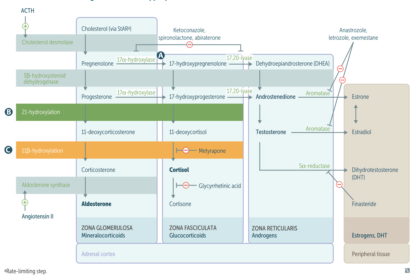
- A stimulus triggers the secretion of CRH, leading to increased secretion of ACTH by the pituitary gland.
- ACTH stimulates the conversion of cholesterol into pregnenolone in the adrenal cortex.
- Enzymatic pathways trigger DHEA and androstenedione production in the zona reticularis.
- Androstenedione is converted into testosterone, which is then transformed into dihydrotestosterone via 5α-reductase.
- Aromatase converts testosterone into estradiol and androstenedione, which is then transformed into estrone.
Mnemonic
Girls’ body are more fragrant…So aromatase converts from andro- to estrogen.
Tip
The testosterone and estrogen precursors DHEA and androstenedione are produced in the adrenal cortex in both male and female individuals. Testosterone is produced by Leydig cells in the testicles and, to a lesser degree, by the theca interstitial cells in the ovaries.
Estrogen
Ovarian estrogen synthesis
Estrogen synthesis primarily takes place in the ovarian granulosa cells.
- LH stimulates androgen synthesis in ovarian theca cells.
- FSH stimulates conversion of androgens to estrogens.
- Conversion of androgens to estrogens is catalyzed by the aromatase enzyme.
Extra-ovarian estrogen synthesis
Estrogen is also produced in other aromatase-containing tissues:
- Fatty tissue
- Placenta
- Testicles
- Adrenal glands
Estrogen types
There are three types of estrogen: estradiol, estrone, and estriol.
- Potency: estradiol > estrone > estriol
- During pregnancy, estrogen concentration increases.
- Estradiol and estrone: 100-fold increase
- Estriol: 1000-fold increase (it is secreted by the placenta as unconjugated estriol and a marker of fetal health)
- Fetal DHEA enters the maternal serum and is converted by the placenta to unconjugated estriol, which is then rapidly conjugated by the liver to a water-soluble form that can be excreted in urine.
Estrogen effects
- Bones: ↑ bone formation by inhibiting bone resorption (induces osteoclast apoptosis)
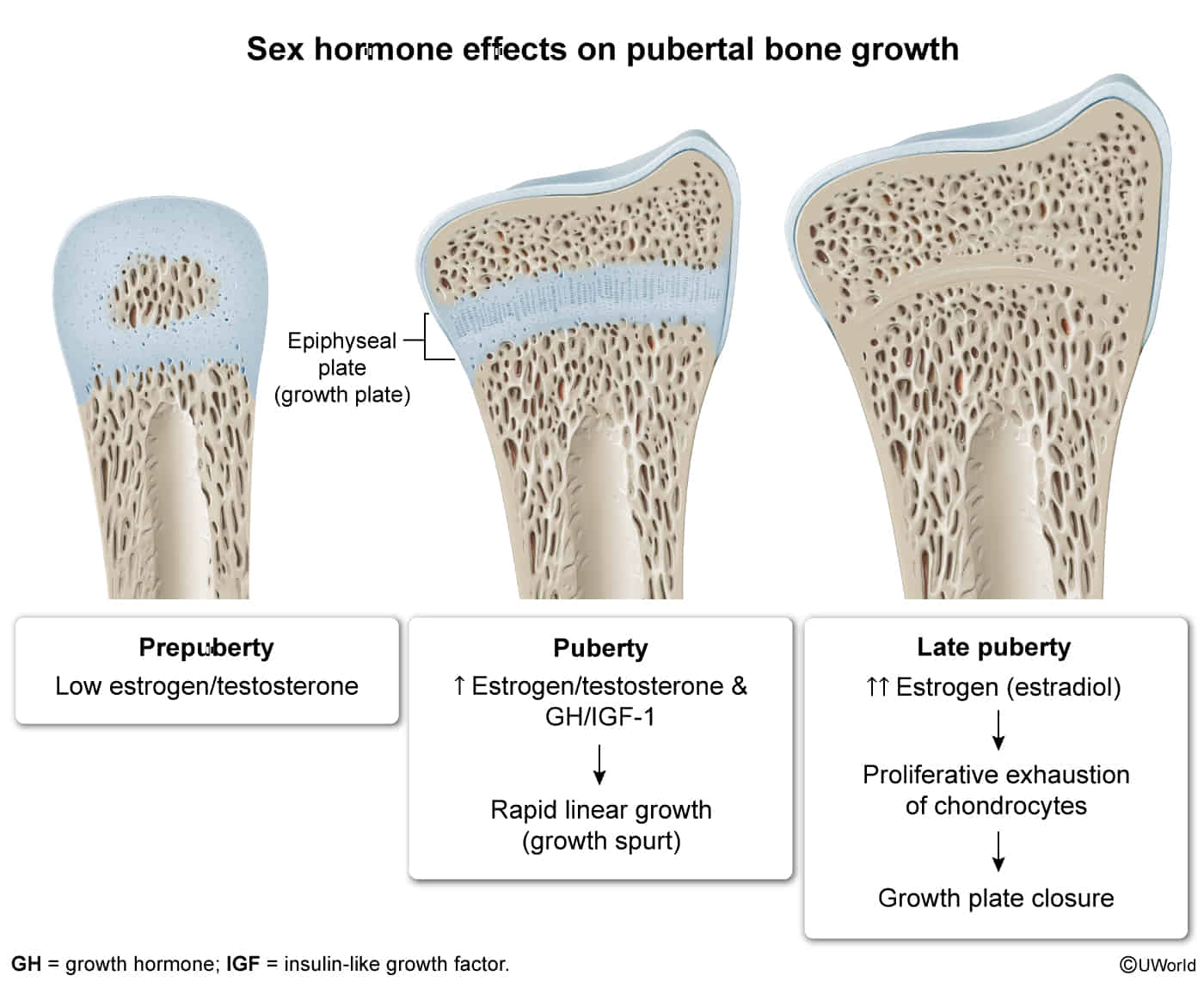
- A growth spurt occurs during early puberty primarily due to positive feedback by estrogen on growth hormone (GH) production. GH stimulates insulin-like growth factor 1 (IGF-1) synthesis, which leads to the differentiation and proliferation of chondrocytes in the epiphyseal plate and therefore increased linear growth
- At high doses, estradiol, a potent estrogen-derived steroid hormone, promotes senescence of chondrocytes and therefore closure of the epiphyseal plate (ie, growth plate). This relates to short stature in idiopathic precocious puberty

- Blood vessels: protective effect against atherosclerosis
- Blood clotting: ↑ risk of thrombosis
- Kidneys: ↑ water and sodium retention (may contribute to edema)
- Protein synthesis
- Slightly ↑ (anabolic effect)
- ↑ Transport proteins (e.g., SHBG, TBG)
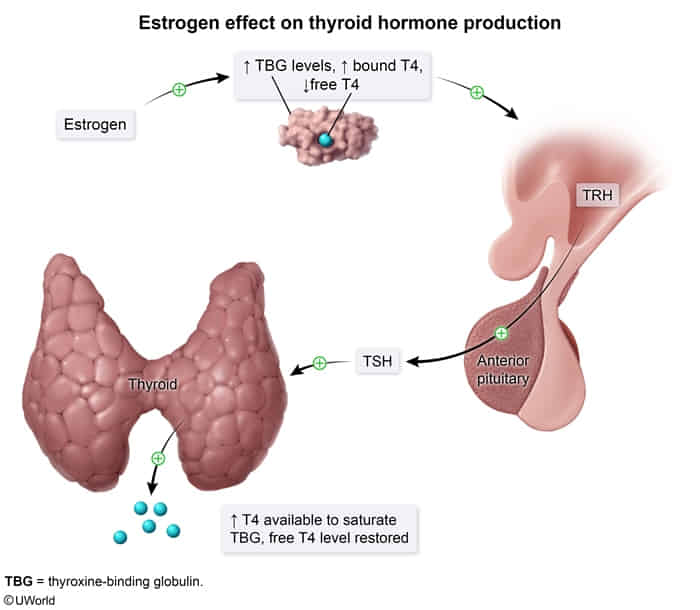
- An increase in estrogen activity, as seen in pregnancy or postmenopausal estrogen replacement therapy, increases the level of thyroxine-binding globulin, causing a corresponding reduction in free T4 and T3 levels. In patients with a normal hypothalamic-pituitary-thyroid axis, this reduction will result in a transient increase in TSH, leading to increased thyroid hormone production until the additional TBG becomes saturated with thyroid hormone and free T4 and T3 levels are restored (ie, patients remain euthyroid). Therefore, an increase in the TBG level leads to an increase in total T4 and T3 (bound plus free fractions).

- An increase in estrogen activity, as seen in pregnancy or postmenopausal estrogen replacement therapy, increases the level of thyroxine-binding globulin, causing a corresponding reduction in free T4 and T3 levels. In patients with a normal hypothalamic-pituitary-thyroid axis, this reduction will result in a transient increase in TSH, leading to increased thyroid hormone production until the additional TBG becomes saturated with thyroid hormone and free T4 and T3 levels are restored (ie, patients remain euthyroid). Therefore, an increase in the TBG level leads to an increase in total T4 and T3 (bound plus free fractions).
- Adipose tissue: female distribution of adipose tissue
- Liver
- ↓ Bilirubin excretion
- ↑ HDL and ↓ LDL
Related drugs
Aromatase inhibitors
- Agents
- Anastrozole
- Letrozole
- Exemestane
- Mechanism of action
- Inhibition of aromatase → ↓ conversion of androstenedione to estrone, ↓ conversion of testosterone to estradiol → ↓ tumor growth
- Indications
- Postmenopausal estrogen receptor-positive breast cancer
- Letrozole: used for ovulation induction (e.g., in PCOS)