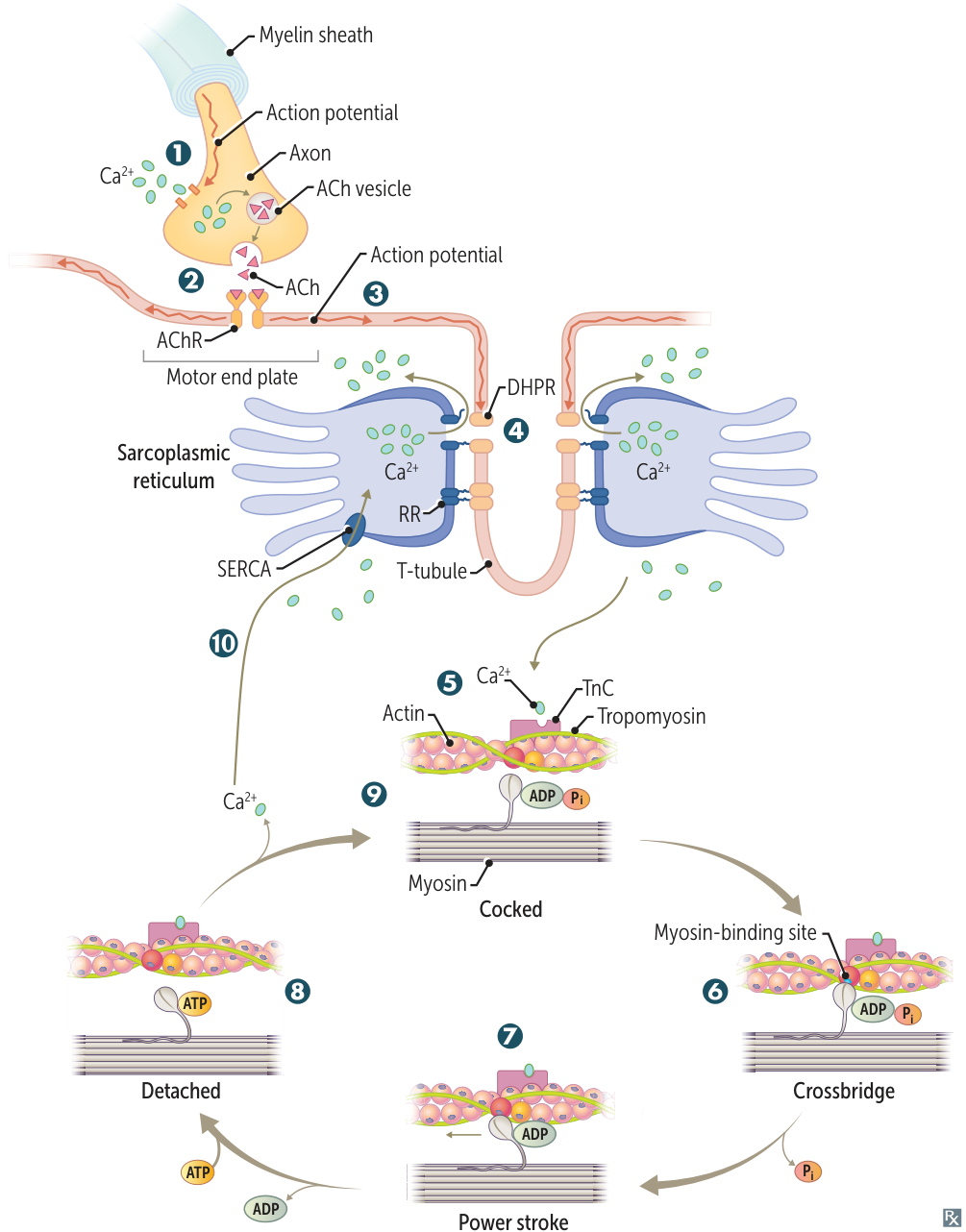
- Action potential opens presynaptic voltage-gated Ca2+ channels, inducing acetylcholine (ACh) release.
- Postsynaptic ACh binding leads to muscle cell depolarization at the motor end plate.
- Depolarization travels over the entire muscle cell and deep into the muscle via the T-tubules.
- Membrane depolarization induces conformational changes in the voltage-sensitive dihydropyridine receptor (DHPR) and its mechanically coupled ryanodine receptor (RR) → Ca2+ release from the sarcoplasmic reticulum (buffered by calsequestrin) into the cytoplasm.
- Tropomyosin is blocking myosin-binding sites on the actin filament. Released Ca2+ binds to troponin C (TnC), shifting tropomyosin to expose the myosin-binding sites.
- Myosin head binds strongly to actin (crossbridge). Pi released, initiating power stroke.
- During the power stroke, force is produced as myosin pulls on the thin filament A. Muscle shortening occurs, with shortening of H and I bands and between Z lines (HI, I’m wearing shortZ). The A band remains the same length (A band is Always the same length). ADP is released at the end of the power stroke.
- Binding of new ATP molecule causes detachment of myosin head from actin filament. Ca2+ is resequestered.
- ATP hydrolysis into ADP and Pi results in myosin head returning to high-energy position (cocked). The myosin head can bind to a new site on actin to form a crossbridge if Ca2+ remains available.
- Reuptake of calcium by sarco(endo)plasmic reticulum Ca2+ ATPase (SERCA) → muscle relaxation.