Only globin present in all major hemoglobins; essential for life
Principal adult hemoglobin chain; most clinically relevant
Almost identical to β but less abundant; forms minor adult HbA₂
Highest O₂ affinity; critical for fetal-maternal O₂ transfer
Hemoglobins
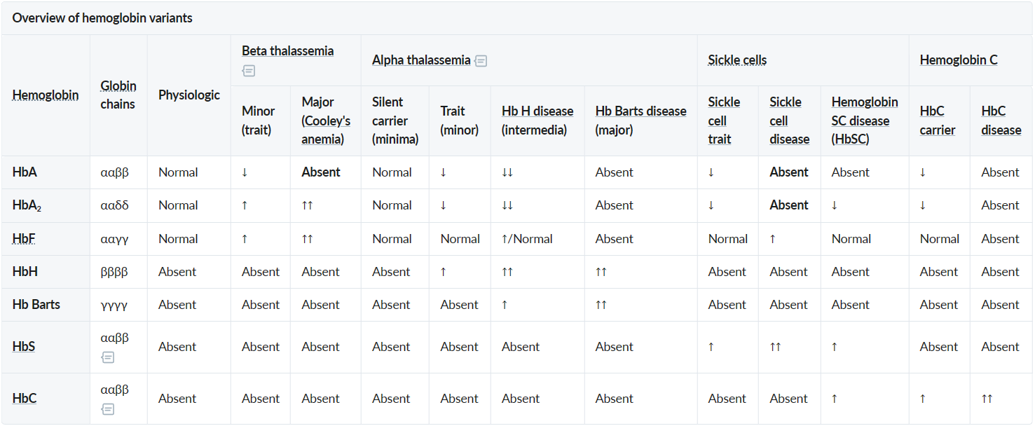
HbF: has significantly higher oxygen affinity than adult hemoglobin A. This allows fetal hemoglobin to extract more oxygen from the mother’s adult hemoglobin in the placenta, providing the developing fetus with an adequate supply of oxygen.
HbS: Point mutation in the β-globin gene resulting in the replacement of glutamic acid by valine
HbC: Point mutation in the β-globin gene resulting in the replacement of glutamic acid by lysine
Hb-electrophoresis
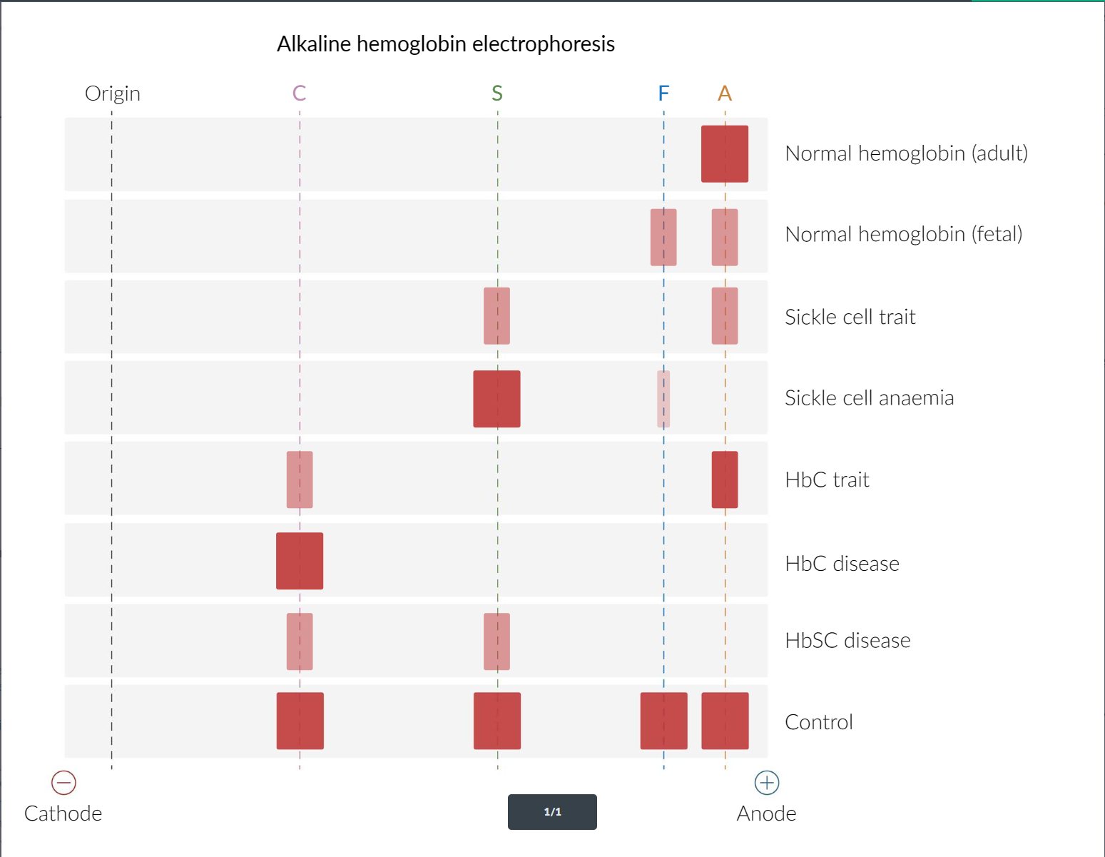
Normal hemoglobin consists primarily of hemoglobin A (HbA), which migrates rapidly toward the positive electrode (anode) because of its negative charge.
Hemoglobin S (HbS) is an abnormal type of hemoglobin in which a nonpolar amino acid (valine) replaces a negatively charged amino acid (glutamate) in the beta globin chain. This amino acid replacement decreases the negative charge on the HbS molecule, which causes HbS to move more slowly toward the anode.
Glutamate (-) → Valine (=)
Similarly, hemoglobin C (HbC) has a glutamate residue replaced by lysine in the beta globin chain. Because lysine is a positively charged amino acid, HbC has even less total negative charge than HbS and moves even more slowly toward the anode. Both HbC and HbS result from missense mutations, a type of mutation in which a single base substitution results in a codon that codes for a different amino acid.