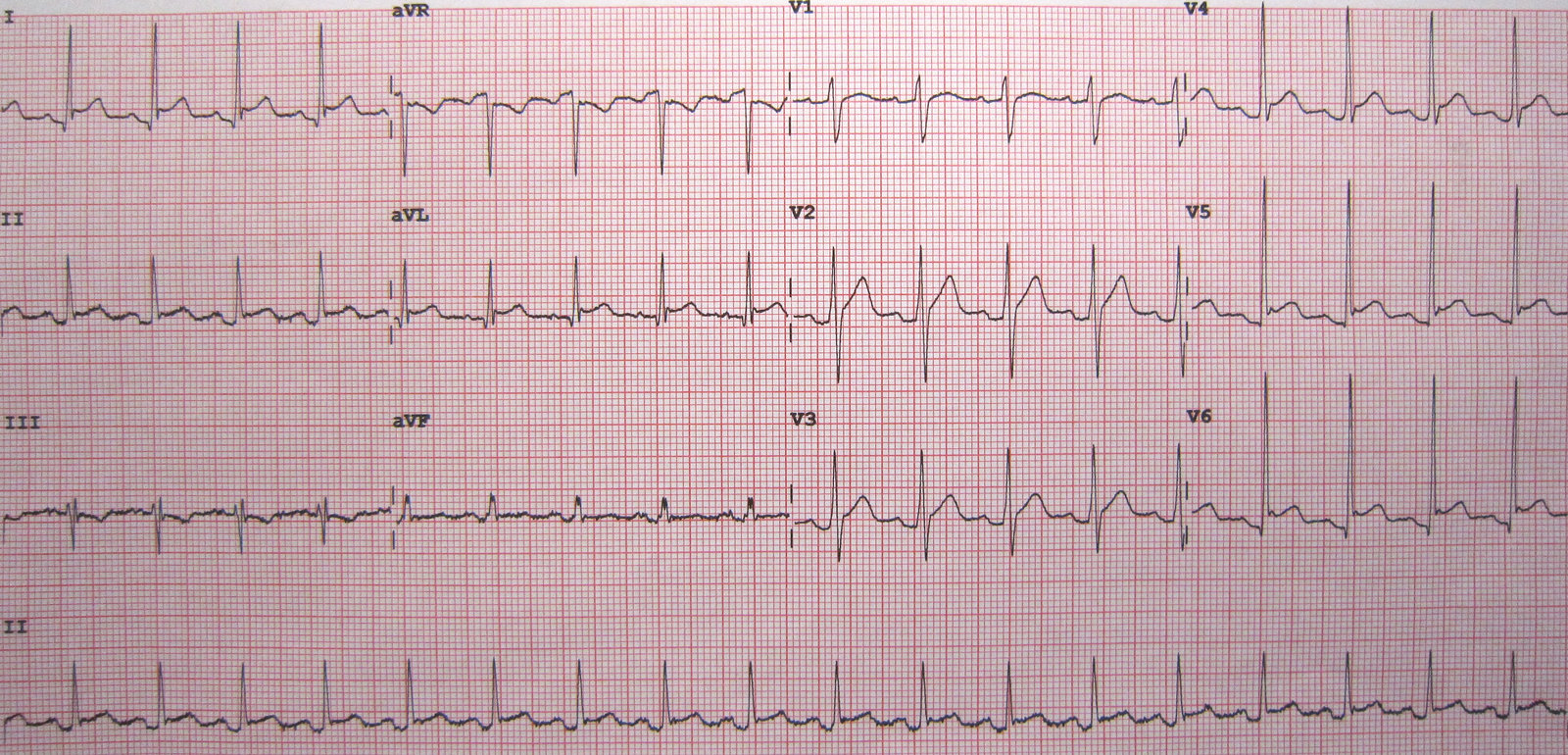
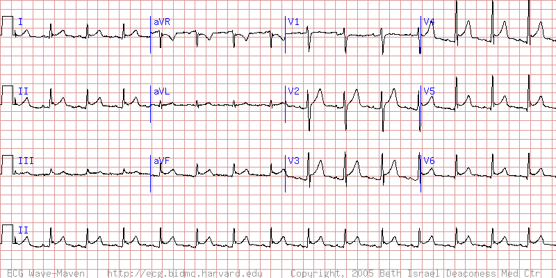
ECG features of pericarditis Not all patients go through all stages and manifestations may vary. In particular, pericarditis due to uremia may not involve characteristic ECG changes. Stage 1: diffuse ST elevations, ST depression in aVR and V1, PR segment depression Stage 2: ST segment normalizes in ∼ 1 week. Stage 3: inverted T waves Stage 4: ECG returns to normal baseline (as prior to onset of pericarditis) after weeks to months.
In contrast to myocardial infarction, pericarditis is characterized by a diffuse distribution of ST elevations on ECG.See also “Differential diagnoses of ST elevations on ECG.”